
Fig. 2.
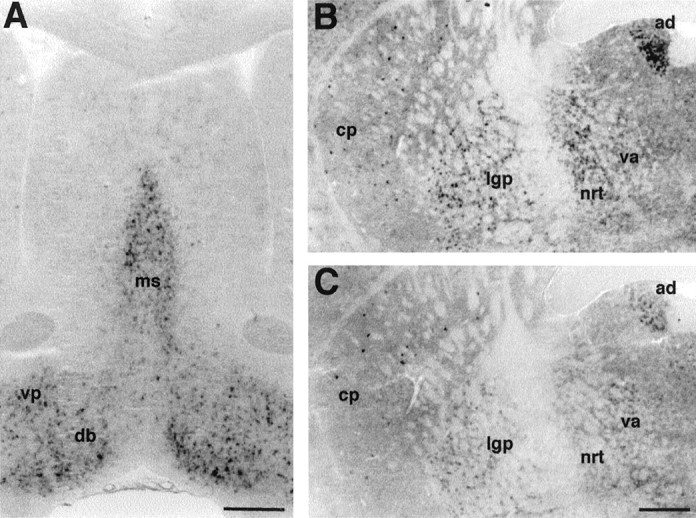
Expression of mHCN2 and mHCN4 mRNA in the mouse basal forebrain. A, Coronal section (bregma, +0.74) showing mHCN2 labeling in the medial septum (ms), vertical and horizontal limb of the diagonal band (db), and ventral pallidum (vp). B, Coronal section (bregma, −0.82) showing staining by the mHCN2 probe of cells in the caudate putamen (cp), lateral globus pallidus (lgp), thalamus nucleus reticularis (nrt), ventral anterior thalamic nucleus (va), and anterodorsal thalamic nucleus (ad).C, Coronal section (bregma, −0.82) showing staining by the mHCN4 probe of cells in the caudate putamen, lateral globus pallidus, and anterodorsal thalamic nucleus. mHCN4 is not present in thalamus nucleus reticularis. Scale bars, 500 μm.