
Fig. 4.
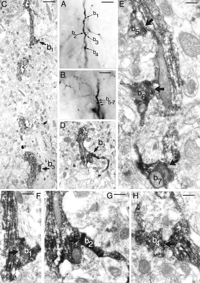
Multiple contacts between CR-IR axons and dendrites. A, B, Two segments of a dendrite (cell 5 in Fig. 2A) are shown at low power. The axon of cell 3 (of Fig. 2) climbs along the dendrite and establishes seven contacts (b1–7) with it, via club-like (b1–4) and “en passant” (b5–7) terminals. C, Low-power electron micrograph of the dendritic segment shown in A, demonstrating the contacts formed by boutons b1and b3. D, The same dendritic segment from another section with bouton b2.E–H, Boutons 1, 2, and 4–7 are shown at higher magnification to form symmetrical synapses (arrows) on the same postsynaptic dendrite (cell 5). The synaptic cleft is not visible in the case of b7 in this section but has been confirmed by goniometer. The accumulation of gold particles in the presynaptic axons and the postsynaptic dendrites clearly indicate their GABA immunoreactivity. However, because of the masking effect of the pre-embedding CR-immunoreaction endproduct, the GABA immunoreactivity is weaker in these elements than in unstained profiles (see Figs. 5,6). Scale bars: A, B, 10 μm; C,D, 1 μm; E, 0.5 μm; F,G, 0.25 μm.