

Abstract
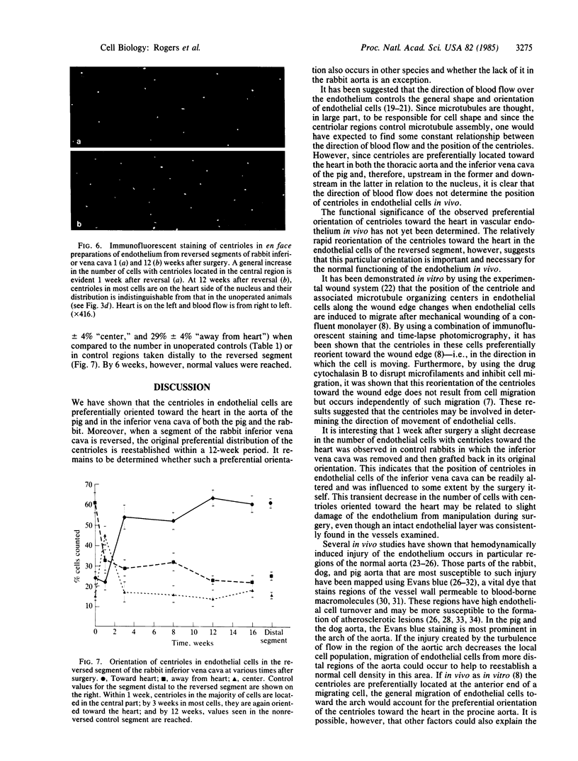
The distribution of centrioles was examined in porcine and rabbit vascular endothelial cells fixed in situ and prepared en face for immunofluorescent staining with rabbit sera that specifically stain these organelles. In endothelial cells lining the major blood vessels of the pig, the centrioles are preferentially located on the heart side of the nucleus regardless of the direction of blood flow. A similar distribution is seen in the inferior vena cava of the rabbit but not in the rabbit aorta. In the major vessels of the pig and in the rabbit inferior vena cava, 60%-80% of the endothelial cells have their centrioles located on the side of the nucleus toward the heart, 10%-20% have them on the side away from the heart, and 7%-15% have them in a central position along the side of the nucleus. To determine whether this preferential orientation is reestablished, microvascular surgical techniques were used to reverse a 3-cm segment of the inferior vena cava between the left renal vein and the common iliac veins of the rabbit. Within 1 week of the reversal, some of the centrioles had migrated from the end away from the heart to a more central position. During the following weeks, an increasing number of endothelial cells had their centrioles located on the heart side of the nucleus; after 12 weeks, values similar to those in the nonreversed inferior vena cava were reached in the reversed segment. The demonstration that the preferential orientation of centrioles on the heart side of the nucleus is reestablished after reversal of a segment suggests that the observed polarity is important for normal functioning of vascular endothelium.
Full text
PDF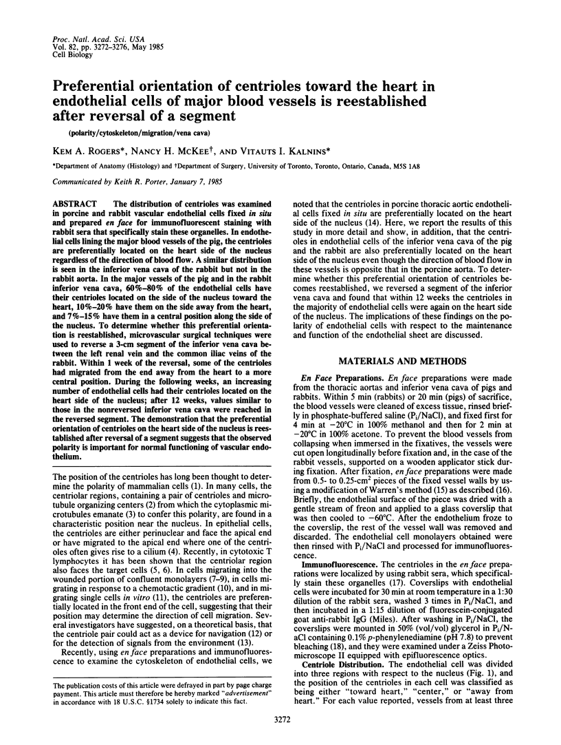


Images in this article


Selected References
These references are in PubMed. This may not be the complete list of references from this article.
- Albrecht-Buehler G., Bushnell A. The orientation of centrioles in migrating 3T3 cells. Exp Cell Res. 1979 Apr;120(1):111–118. doi: 10.1016/0014-4827(79)90542-1. [DOI] [PubMed] [Google Scholar]
- Albrecht-Buehler G. Does the geometric design of centrioles imply their function? Cell Motil. 1981;1(2):237–245. doi: 10.1002/cm.970010206. [DOI] [PubMed] [Google Scholar]
- Bell F. P., Somer J. B., Craig I. H., Schwartz C. J. Patterns of aortic Evans blue uptake in vivo and in vitro. Atherosclerosis. 1972 Nov-Dec;16(3):369–375. doi: 10.1016/0021-9150(72)90084-6. [DOI] [PubMed] [Google Scholar]
- Caplan B. A., Schwartz C. J. Increased endothelial cell turnover in areas of in vivo Evans Blue uptake in the pig aorta. Atherosclerosis. 1973 May-Jun;17(3):401–417. doi: 10.1016/0021-9150(73)90031-2. [DOI] [PubMed] [Google Scholar]
- Connolly J. A., Kalnins V. I. Visualization of centrioles and basal bodies by fluorescent staining with nonimmune rabbit sera. J Cell Biol. 1978 Nov;79(2 Pt 1):526–532. doi: 10.1083/jcb.79.2.526. [DOI] [PMC free article] [PubMed] [Google Scholar]
- Dewey C. F., Jr, Bussolari S. R., Gimbrone M. A., Jr, Davies P. F. The dynamic response of vascular endothelial cells to fluid shear stress. J Biomech Eng. 1981 Aug;103(3):177–185. doi: 10.1115/1.3138276. [DOI] [PubMed] [Google Scholar]
- Flaherty J. T., Pierce J. E., Ferrans V. J., Patel D. J., Tucker W. K., Fry D. L. Endothelial nuclear patterns in the canine arterial tree with particular reference to hemodynamic events. Circ Res. 1972 Jan;30(1):23–33. doi: 10.1161/01.res.30.1.23. [DOI] [PubMed] [Google Scholar]
- Geiger B., Rosen D., Berke G. Spatial relationships of microtubule-organizing centers and the contact area of cytotoxic T lymphocytes and target cells. J Cell Biol. 1982 Oct;95(1):137–143. doi: 10.1083/jcb.95.1.137. [DOI] [PMC free article] [PubMed] [Google Scholar]
- Gerrity R. G., Richardson M., Caplan B. A., Cade J. F., Hirsh J., Schwartz C. J. Endotoxin-induced vascular endothelial injury and repair. II. Focal injury, en face morphology, (3H)thymidine uptake and circulating endothelial cells in the dog. Exp Mol Pathol. 1976 Feb;24(1):59–69. doi: 10.1016/0014-4800(76)90057-5. [DOI] [PubMed] [Google Scholar]
- Gotlieb A. I., May L. M., Subrahmanyan L., Kalnins V. I. Distribution of microtubule organizing centers in migrating sheets of endothelial cells. J Cell Biol. 1981 Nov;91(2 Pt 1):589–594. doi: 10.1083/jcb.91.2.589. [DOI] [PMC free article] [PubMed] [Google Scholar]
- Gotlieb A. I., Spector W. Migration into an in vitro experimental wound: a comparison of porcine aortic endothelial and smooth muscle cells and the effect of culture irradiation. Am J Pathol. 1981 May;103(2):271–282. [PMC free article] [PubMed] [Google Scholar]
- Gotlieb A. I., Subrahmanyan L., Kalnins V. I. Microtubule-organizing centers and cell migration: effect of inhibition of migration and microtubule disruption in endothelial cells. J Cell Biol. 1983 May;96(5):1266–1272. doi: 10.1083/jcb.96.5.1266. [DOI] [PMC free article] [PubMed] [Google Scholar]
- Gutstein W. H., Farrell G. A., Armellini C. Blood flow disturbance and endothelial cell injury in preatherosclerotic swine. Lab Invest. 1973 Aug;29(2):134–149. [PubMed] [Google Scholar]
- Johnson G. D., Nogueira Araujo G. M. A simple method of reducing the fading of immunofluorescence during microscopy. J Immunol Methods. 1981;43(3):349–350. doi: 10.1016/0022-1759(81)90183-6. [DOI] [PubMed] [Google Scholar]
- Kupfer A., Dennert G., Singer S. J. Polarization of the Golgi apparatus and the microtubule-organizing center within cloned natural killer cells bound to their targets. Proc Natl Acad Sci U S A. 1983 Dec;80(23):7224–7228. doi: 10.1073/pnas.80.23.7224. [DOI] [PMC free article] [PubMed] [Google Scholar]
- Kupfer A., Louvard D., Singer S. J. Polarization of the Golgi apparatus and the microtubule-organizing center in cultured fibroblasts at the edge of an experimental wound. Proc Natl Acad Sci U S A. 1982 Apr;79(8):2603–2607. doi: 10.1073/pnas.79.8.2603. [DOI] [PMC free article] [PubMed] [Google Scholar]
- Langille B. L., Adamson S. L. Relationship between blood flow direction and endothelial cell orientation at arterial branch sites in rabbits and mice. Circ Res. 1981 Apr;48(4):481–488. doi: 10.1161/01.res.48.4.481. [DOI] [PubMed] [Google Scholar]
- Malech H. L., Root R. K., Gallin J. I. Structural analysis of human neutrophil migration. Centriole, microtubule, and microfilament orientation and function during chemotaxis. J Cell Biol. 1977 Dec;75(3):666–693. doi: 10.1083/jcb.75.3.666. [DOI] [PMC free article] [PubMed] [Google Scholar]
- McGILL H. C., Jr, GEER J. C., HOLMAN R. L. Sites of vascular vulnerability in dogs demonstrated by Evans blue. AMA Arch Pathol. 1957 Sep;64(3):303–311. [PubMed] [Google Scholar]
- Packham M. A., Rowsell H. C., Jorgensen L., Mustard J. F. Localized protein accumulation in the wall of the aorta. Exp Mol Pathol. 1967 Oct;7(2):214–232. doi: 10.1016/0014-4800(67)90031-7. [DOI] [PubMed] [Google Scholar]
- Reidy M. A., Bowyer D. E. Scanning electron microscopy of arteries. The morphology of aortic endothelium in haemodynamically stressed areas associated with branches. Atherosclerosis. 1977 Feb;26(2):181–194. doi: 10.1016/0021-9150(77)90101-0. [DOI] [PubMed] [Google Scholar]
- Rogers K. A., Kalnins V. I. A method for examining the endothelial cytoskeleton in situ using immunofluorescence. J Histochem Cytochem. 1983 Nov;31(11):1317–1320. doi: 10.1177/31.11.6352798. [DOI] [PubMed] [Google Scholar]
- Rogers K. A., Kalnins V. I. Comparison of the cytoskeleton in aortic endothelial cells in situ and in vitro. Lab Invest. 1983 Dec;49(6):650–654. [PubMed] [Google Scholar]
- Ross R., Glomset J. A. The pathogenesis of atherosclerosis (first of two parts). N Engl J Med. 1976 Aug 12;295(7):369–377. doi: 10.1056/NEJM197608122950707. [DOI] [PubMed] [Google Scholar]
- Ross R., Glomset J. A. The pathogenesis of atherosclerosis (second of two parts). N Engl J Med. 1976 Aug 19;295(8):420–425. doi: 10.1056/NEJM197608192950805. [DOI] [PubMed] [Google Scholar]
- Somer J. B., Schwartz C. J. Focal 3 H-cholesterol uptake in the pig aorta. Atherosclerosis. 1971 May-Jun;13(3):293–304. doi: 10.1016/0021-9150(71)90073-6. [DOI] [PubMed] [Google Scholar]
- Svendsen E., Jørgensen L. Focal "spontaneous" alterations and loss of endothelial cells in rabbit aorta. Acta Pathol Microbiol Scand A. 1978 Jan;86(1):1–13. doi: 10.1111/j.1699-0463.1978.tb02005.x. [DOI] [PubMed] [Google Scholar]
- Warren B. A. A method for the production of "en face" preparations one cell in thickness. J R Microsc Soc. 1965 Dec;85(4):407–413. doi: 10.1111/j.1365-2818.1965.tb02141.x. [DOI] [PubMed] [Google Scholar]