
Figure 2.
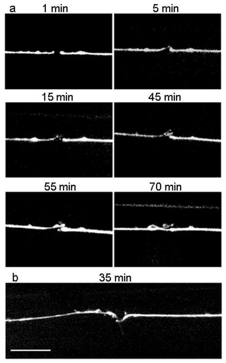
Time-lapse imaging of axonal recovery on-a-chip. (a) Fluorescence images of an ALM neuron at 1, 5, 15, 45, 55 and 70 minutes after the axotomy, respectively. Distal ends are on the left side of the pictures, proximal ones on the right. After 5 minutes, the distal end displays a growth cone that is visible above the proximal stump. At 15 minutes, the growth cone branches off into two. At 45 minutes, a third branch sprouts close to the distal stump. The other two growth cones are out of focus. At 55 minutes, the third branch recesses and the first two branches develop into a broad growth cone. The proximal end starts regrowing. At 70 minutes, the proximal end regrew and reconnected to the distal end a bit further past the distal stump. (b) Fluorescence image of axonal recovery in a different axon 35 minutes after axotomy. The distal growth was misguided and developed sideways (pointing downward on the picture). The proximal end is regrowing towards the distal part. Scale bar is 10 μm.