
Figure 3.
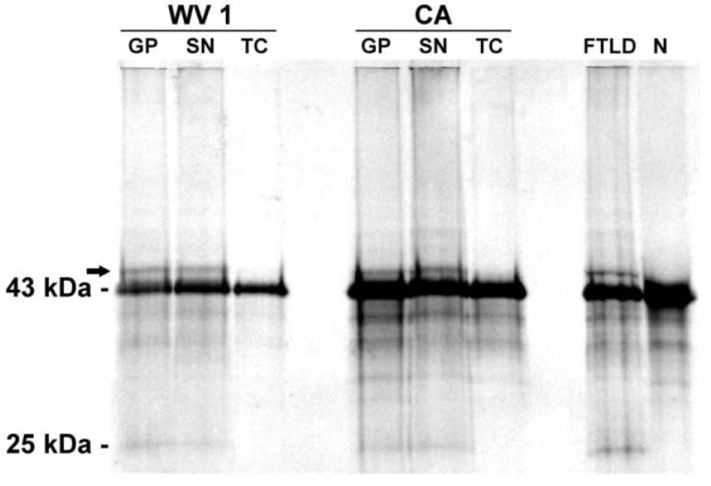
Western blot of Perry patients from WV 1 and CA, one patient with FTLD-U (FTLD) and one neurologically normal control (N) shows the normal 43 kDa TDP-43 band in all subjects. In Perry syndrome, pathologic 25 kDa fragment and hyperphosphorylated (arrow) TDP-43 bands are present in the globus pallidus (GP) and substantia nigra (SN), but not in the temporal cortex (TC). Abnormal bands are similar in Perry patients and FTLD-U, and absent in the control subject.