

Abstract
Adiponectin is a protein hormone involved in maintaining energy homeostasis in metabolically active tissues. It enhances glucose and lipid metabolism via activation of AMP-dependent kinase (AMPK) in skeletal muscle and liver. Energy homeostasis is vital for the heart to work as a pump. In this study, we investigated whether adiponectin and its receptors are expressed in adult ventricular cardiomyocytes. We observed adiponectin transcript and protein in cultured ventricular cardiomyocytes isolated from adult rat, by quantitative real-time PCR, ELISA assays, Western blots, and immunofluorescent staining. In addition, we detected adiponectin receptor (AdipoR1 and AdipoR2) expression in the heart. AdipoR1 was expressed in rat myocardium at a level of about 50% of that in skeletal muscle; whereas adipoR2 was expressed at a similar level to that in liver. Rosiglitazone, a Peroxisome proliferator activated receptor γ (PPARγ) activator, substantially elevated expression of adiponectin in cultured cardiomyocytes and its secretion into cultured media. Rosiglitazone also increased adipoR1 and adipoR2 expression in cardiomyocytes. Treatment of recombinant globular adiponectin in cultured cardiomyocytes increased fatty acid oxidation and glucose uptake via activation of AMPK, suggesting a role for adiponectin in cardiac energy metabolism. Together, these data establish the existence of a local cardiac-specific adiponectin system that is regulated by PPARγ. Moreover, these findings indicate a role for adiponectin on normal myocardial energy homeostasis, in part, through the activation of AMPK.
Keywords: adiponectin, adipoR1, adipoR2, rosiglitazone, AMPK, Fatty acid oxidation, glucose uptake, cardiomyocytes
Introduction
Adiponectin is a protein hormone secreted predominantly by differentiated adipocytes and involved in energy homeostasis. It modulates glucose and lipid metabolism via activating AMP-activated protein kinase (AMPK) activities [1, 2]. Adiponectin contains an N-terminal collagenous domain and a C-terminal globular domain. The globular domain as a proteolytic product is the most potent form that activates AMPK activity, enhances fatty acid oxidation (FAO) and glucose uptake in skeletal muscle and liver [2]. The adiponectin receptors, AdipoR1 and AdipoR2, have been characterized [3]. AdipoR1 is abundantly expressed in skeletal muscle, whereas AdipoR2 is predominantly expressed in the liver [3]. Adiponectin and its receptor can be detected in cultured atrial cardiomyocytes [4]. Moreover, left-ventricular pressure-overload induced by transverse aortic banding in adiponectin-deficient mice results in exacerbated concentric cardiac hypertrophy and increased mortality with increased extracellular signal-regulated kinase (ERK) and diminished AMPK signaling in mouse heart [5]. However, it is uncertain whether adiponectin and/or its receptors are expressed in adult ventricular cardiomyocytes and how they are regulated.
Fatty acids and glucose are essential fuel sources utilized by cardiac muscle to meet its energy needs. Peroxisome proliferator activated receptors (PPARs: α, β/δ and γ), members of the nuclear hormone receptor superfamily of ligand-dependent transcription factors, transcriptionally regulate energy metabolism and homeostasis; whereas AMPK post-transcriptionally activates glucose and fatty acid metabolism during hypoxic stress or extreme metabolic demand [6–8]. However, little is known about the upstream regulator(s) that modulate AMPK activities in cardiomyocytes. As important determinants of energy metabolism; PPARγ, adiponectin and AMPK may play important roles in maintaining energy homeostasis in cardiomyocytes. Thus, understanding their role in cardiomyocyte function is crucial.
In the present study, we demonstrate that adiponectin and its receptors are expressed and relatively abundant in adult ventricular cardiomyocytes. PPARγ activation enhances the expressions of adiponectin and its receptors. Moreover, adiponectin treatment activates AMPK and elevates FAO and glucose uptake in cultured ventricular cardiomyocytes. These results establish the importance of the adiponectin system in adult ventricles and implicate an important role for adiponectin in maintaining myocardial energy homeostasis.
Materials and Methods
1.1. Culture of adult rat cardiomyocytes
Adult rat (Sprague Dawley, Taconic, Petersburgh, NY) ventricular cardiomyocytes were isolated and cultured as described previously [9] with minor modifications. Adult rats (2–3 month-old) were anesthetized with ketamine (4 mg/kg IM) and xylazine (50 mg/kg IM), as previously described [10]. The heart was quickly removed and retrogradely perfused under constant pressure (60 mm Hg) at 37°C for ~4 min with a Ca2+-free buffer containing (in mM) (130)NaCl, (5)KCl, (5)MgSO4, (21)glucose, (1)NaH2PO4, (20)taurine, (5)creatine, (5)Na pyruvate, (23)HEPES. Enzymatic digestion was initiated by adding collagenase II (1 mg/ml, Worthington) and CaCl2 (50 μM) to the perfusion solution. After ~10 min of digestion, the ventricle was quickly removed and gently teased into small pieces with fine forceps in the perfusion solution with 50 μM CaCl2. The ventricle tissues were further dissociated using plastic transfer pipettes with different sized openings (2-mm, 1.5-mm, and then 1-mm diameters) until all the large pieces were dispersed into cell suspension. The solution was filter with 70 μm nylon cell strainer (Fisher Scientific, Waltham, MA) to exclude un-digested tissues. After the cardiomyocytes were pelleted by gravity for ~10 min, the supernatant was aspirated and the cardiomyocytes were resuspended in the perfusion solution containing serum free and calcium (50 μM). The above steps were repeated 3 times to eliminate other cell types such as adipocytes. Increased calcium concentrations were achieved by gradually adding CaCl2 from 50 mM to 1 mM. The cardiomyocytes pooled from three hearts were then plated in six culture dishes which were pre-coated for 1 hour with 10 μg/ml mouse laminin (Invitrogen, Carlsbad, CA) in PBS at 37°C. Cardiomyocytes were cultured for 1 hour to allow attachment to the culture dish and carefully washed with serum free M199 media 3 times. The un-attached cells were discarded. The above cardiomyocytes were cultured for 12 hours in M199 media with 5% fetal bovine serum, penicillin (100 U/mL) and streptomycin (100 μg/mL). After 24 hours of culturing in serum-free conditions, the cells either collected for transcript and protein analyses or for further experiment with rosiglitazone (10 μM) treatments. Potential contamination of adipocytes was further excluded by negative Oil-red O staining on the above cultured cardiomyocytes. The culture protocol yielded an average of 80% rod-shaped myocytes at a plating density of 50 cells/mm2 that were viable at pH 7.2 for 48hr. Experiments were performed the day following isolation and culture.
All experiments were conducted in accordance with the principles and procedures described by NIH Guidelines for the Care and Use of Experimental Animals and were approved by Institutional Animal Care and Use committee of Atlanta University Center.
1.2. Culture of 3T3-L1 preadipocytes and differentiated adipocyte
3T3-L1 preadipocytes were cultured in DMEM containing 10% calf serum with penicillin (100 U/mL) and streptomycin (100 μg/mL) [11]. For MDI (methylisobutylxanthine, dexamethasone, and insulin) induced differentiation, confluent preadipocyte monolayers were incubated for 48 h in DMEM containing 10% fetal bovine serum and a differentiation cocktail consisting of methylisobutylxanthine (115 μg/mL), dexamethasone (390 ng/mL), and insulin (10 μM). After 48 h, the cells were maintained in DMEM containing 10% fetal bovine serum, antibiotics, and insulin. The medium was changed every 2 days. The cell morphology was monitored daily for the appearance of cytoplasmic lipid droplets, using a phase-contrast microscope and oil red O staining.
1.3. Transcript analyses
RNA isolation
The left ventricle, liver and skeletal muscle (tibialis anterior muscle) from anesthetized rats were harvested and frozen in liquid nitrogen. After homogenization, total RNA from the above tissues was isolated using the RNA isolation kit (Qiagen, Valencia, CA) per the manufacturer’s instructions. Total RNA was also extracted from cell lysates from cardiomyocytes and adipocytes using the same method and reverse transcribed into cDNA using the Superscript first strand cDNA synthesis kit (Invitrogen, Carlsbad, CA) per manufacturer’s instructions.
Quantitative Real-time PCR
The mRNA levels were quantitated by using quantitative real-time PCR (QPCR). Specific primers were designed for each gene of interest followed by standard PCR reaction chemistry with the addition of the fluorescent DNA-binding dye, SYBR Green I (Roche, Indianapolis, IN). Serial dilutions of an external standard with a predefined known concentration were used to create a standard curve. QPCR results for each gene were analyzed and reported by LightCycler software version 3.5 (Roche, Indianapolis, IN). The determination of unknown sample concentration involved determination of the crossing point value, which correlated inversely with the log of the initial template concentration. Values were controlled for reverse transcription efficiency and cDNA loading by normalizing with an endogenous control (β-actin).
Primer sequences (all for rat): adipocyte fatty acid–binding protein (aP2, GenBank accession: NM_053365), forward: TTGTGGGGACCTGGAAACT, reverse: TGTCATCTGGGGTGATTTCA (222 bp); Adiponectin (GenBank Accession No. NM_144744), forward: 5′-TCCTGGTCACAATGGGATACC-3′, reverse: 5′-ATCTCCTGGGTCACCCTTAGG-3′ (109 bp)[12]; AdipoR1 (GenBank accession: DQ148391), forward: 5′-GCTGGCCTTTATGCTGCTCG-3′, reverse: 5′-TCTAGGCCGTAACGGAATTC-3′ (158bp); AdipoR2 (GenBank accession: DQ148392), forward: 5′-CCACAACCTTGCTTCATCTA-3′, reverse: 5′-GATACTGAGGGGTGGCAAAC-3′ (100bp). The PCR amplification reaction was done in 20 μl containing 4 mM MgCl2, 0.5 μM each primer pair, 2 μl FastStart SYBR Green mix (Roche, Indianapolis, IN), and 2 μl template cDNA. Efficiency for each primer pair was assessed by using serial dilutions of RT product.
1.4. Protein analyses
Western blots
For the detection of adiponectin, total protein samples were extracted and subjected to SDS-PAGE electrophoresis. For the detection of adipoR1 and adipoR2, protein samples from both plasma membrane (PM) and light microsome (LM) fractions were extracted according to the method of Mitsumoto and Klip [13]. Briefly, the heart, liver and skeletal muscle (about 30 mg) were homogenized in homogenization buffer A (250 mmol/l sucrose, 5 mmol/l NaN3, 2 mmol/l EGTA, 200 μmol/l phenylmethylsulfonyl fluoride [PMSF], 1 μmol/l pepstatin A, 1 μmol/l aprotinin, and 20 mmol/l HEPES [pH 7.4]). The homogenate was centrifuged at 760 x g for 5 min to remove nuclei and unbroken cells. The supernatant was centrifuged at 31,000 x g for 60 min to pellet the crude plasma membrane (PM). The light microsomes (LM) were collected from the 31,000 x g supernatant by centrifugation at 190,000 x g for 60 min (Optima TLX Utracentrifuge, Beckman, Fullerton, CA). Both PM and LM pellets were suspended in the homogenization buffer and frozen at −80°C until use. For the detection of adiponectin, protein samples were extracted from isolated adult rat cardiomyocytes. Approximately 30–50mg isolated adult rat cardiomyocyte was homogenized in buffer B (50 mM TRIS-HCl, containing 105mM NaCl, 1% NP-40, 1% sodium deoxycholate, 0.1% SDS and 2mM EDTA, 5 mmol/l NaN3, 2 mmol/l EGTA, 200 μmol/l phenylmethylsulfonyl fluoride [PMSF], 1 μmol/l pepstatin A, 1 μmol/l aprotinin (pH=7.5) and centrifuged (14 000 rpm) for 15 minutes. The supernatant was collected as a cytosol fraction. For immunoblotting, 50 μg of denatured protein for detecting adiponectin and 30 μg of denatured protein for detecting adipoR1, adipoR2 were subjected to sodium dodecyl sulfate-polyacrylamide gel electrophoresis (SDS-PAGE) on a 12.5% polyacrylamide gel (Bio-Rad, Hercules, CA), and separated proteins were electrophoretically transferred onto polyvinylidene difluoride (PVDF) membranes (Bio-Rad, Hercules, CA). Nonspecific binding was blocked by incubation with 5% nonfat dry milk for 1 hour. The PVDF membranes were then incubated with the following antibodies overnight at 4°C. Rabbit antibodies for adipoR1 (Alpha Diagnostic, San Antonio, TX) as well as adipoR2 (Phoenix Pharmaceuticals, Burlingame, CA) were used. Both of these antibodies have been used in previous studies for the specific detection of adiponectin receptors [14, 15]. Polyclonal antibody for adiponectin was obtained from Santa Cruz biotechnology (Santa Cruz, CA, cat#: sc-17044-R, lot #:I1304). A blocking peptide (Santa Cruz Biotechnology, Santa Cruz, CA, cat#: sc-17044-P, lot #:H142) was also obtained and applied as a negative control. The concentration of anti-adiponectin antibody was 1:1000. After washing in Tween-20 buffer, the membranes were incubated with goat anti-rabbit horseradish peroxidase (1:1000) (Santa Cruz Biotechnology, Santa Cruz, CA) for 1 hour at room temperature. Antibodies for acetyl CoA carboxylase II (ACC) and phosphorylated ACC (pACC) were purchased from Santa Cruz Biotechnology. The Typhoon phosphoImager scanner (Amersham, Piscataway, NJ) was used for quantitative measurement of protein signals. Sample loadings were normalized by immunoblotting with an anti-actin polyclonal antibody (SIGMA-Aldrich, St. Louis, MO).
ELISA assessment of adiponectin concentration in media of cultured cardiomyocytes
Media from adult cardiomyocyte cultures were analyzed using a Rat Adiponectin ELISA kit from B-Bridge (Sunnyvale, CA). The assay was performed according to the manufacturer’s protocol. Briefly, the wells of a microtiter plate coated with a pretitered amount of adiponectin antibody were loaded with 100 μl volumes of duplicate samples and adiponectin standards in the order of ascending concentration. After a second biotinylated anti-rat polyclonal antibody was added and washed, 100 μl of enzyme solution (streptavidin-horseradish peroxidase) and 100 μl of substrate solution were added. The enzyme activity was measured spectrophotometrically by the increased absorbance at 450 nm. The amount of captured adiponectin in the samples was calculated from a reference curve generated in the same assay with reference standards of known concentrations of adiponectin with SOFTmax PRO software (Molecular Devices Corporation, Sunnyvale, CA). The minimum detectable limit is 15.6pg/ml of adiponectin.
1.5. Indirect immunofluorescent staining
Expression and localization of adiponectin, adipoR1 and adipoR2 in cardiomyocytes were examined using indirect fluorescent staining. Cultured rat adult cardiomyocytes in chamber slides were fixed in 4% formalin buffer for 20 min. After serial washing with PBS and blocking in 10% horse serum in PBS for 1 hour, the same primary antibodies for adiponectin, adipoR1 and adipoR2 used for immunoblotting were diluted in 10% FBS/PBS and applied to the slides and incubated for 12 hours at 4°C. Secondary anti-IgG antibody of the specific species of the primary antibodies and conjugate with Alexa Fluor dyes (Molecular Probe, Carlsbad, CA) were used. Cardiomyocytes treated with normal rabbit IgG served as negative control. The immunofluorescent staining cardiomyocytes were then examined using a confocal microscope.
1.6. 14C Palmitate oxidation assay
Rates of FAO in cultured cardiomyocytes on laminin-coated plates were evaluated by 14C Palmitate oxidation assays. Measurements of cellular palmitate oxidation rates were performed as described [16, 17]. Cultured cardiomyocytes from 2 month-old rats treated with adiponectin (2 μg/ml) were pre-incubated in assay buffer containing [(in mMol/L), HEPES (20), NaH2PO4 (1), MgSO4 (0.4), CaCl2 (1), NaCl (120), KCl (5), glucose (5), palmitic acid (0.2), oleic acid (0.2), pH 7.4] for 30 min at 37°C. Subsequently, a trace amount of [1-14C]palmitic acid (1μCi/ml, PerkinElmer life Sciences, Waltham, MA) was added and the pre-incubation was continued for an additional 30 min in order to reach steady state. Thereafter, a vial containing 500 μl of CO2-trapping medium (NaOH, 0.1 Mol/L) was inserted and the flasks were subsequently sealed airtight. Oxidation was terminated immediately after 30 min by injection of 500 μl HClO4 (5 Mol/L) through the seal on the lid of the flask. Flasks were then stored at 4°C overnight and the trapping medium was assessed for 14C activity by liquid scintillation counting.
1.7. Glucose uptakes in isolated cardiomyocytes
Glucose transport assays were performed in triplicate in 12-well (22 mm diameter) laminin-coated tissue culture plates as described before [17]. Laminin-plated cardiomyocytes from 2 month-old rats were washed 3 times with PBS and cultured in 1 ml of glucose- and serum-free DMEM. Then globular adiponectin (2 μg/ml) and insulin (10nMol/L) were added. After 30 minutes, 1 μCi/μl [3H] 2-deoxyglucose (PerkinElmer life Sciences, Waltham, MA) was added to the culture dish. After 30 minutes, the cultured cardiomyocytes were washed 3 times with cold PBS and lysed in 500 μl of NaOH 1N for 20 minutes at 37°C. A 400-μl aliquot of lysed cells was counted to determine the specific activity of [3H] 2-deoxyglucose normalized to protein concentration.
1.8. Myocardial Malonyl CoA content
Malonyl CoA content was measured by HPLC in cardiomyocytes with and without treatment of globular adiponectin (2 μg/ml) for 30 minutes. Malonyl-CoA was extracted from cultured cardiomyocytes with 5% sulfosalicylic acid containing 50 μM of dithioerythritol in 1:9 w/v (mg/μl) proportion and measured with HPLC separation using previously described methodology [18, 19].
1.9. Malonyl CoA decarboxylase (MCD) activity
The enzyme activity was assayed according to a published method [20] with slight modification. MCD activity were estimated by measuring the amount of 14CO2 generated from [2-14C] malonyl-CoA in cardiomyocytes with and without treatment of globular adiponectin (2 μg/ml) for 30 minutes. The cell lysis buffer containing 75 mM KCl, 20 mM sucrose, 10 mM Hepes, 1 mM EGTA, 50 mM NaF, 5 mM NaPPi, 1 mM dithiothreitol, and a protease inhibitor cocktail was used. Samples were subjected to sonication on ice for ~5 s and whole cell lysates were used. Protein concentrations of the cell lysates were determined to normalize the final results. The reaction mixture containing 10 μmol of the Tris-HCl buffer (pH 8.0), 0.01 μmol of DTE, 0.02 μmol of [2-14C] malonyl-CoA (Amersham, Piscataway, NJ), and the enzyme in a total volume of 0.1 ml was incubated for 10 min at 30°C. The 14CO2 generated was assessed as described in palmitate oxidation measurement.
2.0. Statistical Analysis
Comparisons were analyzed by Student’s t test (for two groups; P<0.05) or one factor or mixed, two-factor analysis of Variance (ANOVA) followed by Student-Newman-Keuls and Bonferroni tests (for three or more groups). Results are presented as mean±SEM.
Results
Adiponectin is expressed in cardiomyocytes isolated from adult left ventricles from rats
Adiponectin is an important regulator of energy metabolism as indicated by its potent effects on FAO and glucose utilization in skeletal muscle and in liver [2]. QPCR revealed that adiponectin mRNA was expressed in RNA samples isolated from adult rat cardiomyocytes, albeit at modest levels compared to differentiated adipocytes (Figure 1a). Adiponectin transcript was also detectable in cultured neonatal cardiomyocytes but at a much lower concentration than that of adult cardiomyocytes (data not shown). On the other hand, we did not detect any adiponectin expression in other cell types such as HeLa cells, predifferentiated 3T3-L1 cells and human embryonic kidney-293 (HEK-293) cells (data not shown). To assess whether the cardiomyocyte cultures were contaminated with adipocytes, we examined the expression of adipocyte fatty acid-binding protein, aP2, an adipocyte marker, in cultured cardiomyocytes and differentiated adipocytes with RT-PCR. While aP2 was not detected in RNA samples from adult cardiomyocyte, the expression of aP2 was abundant in RNA samples from the differentiated adipocytes (Figure 1b). Therefore, the adiponectin transcript detected from cultured cardiomyocytes is not likely from adipocyte contamination. Western blots using adiponectin-specific antibody for adiponectin revealed that adiponectin protein is expressed in rat adult cardiomyocytes, but largely undetectable in neonatal rat cardiomyocytes (Figure 1c). Preincubation of the protein samples with a blocking epitope peptide largely eliminated the specific band of adiponectin in protein samples from both cardiomyocytes and adipocytes (Figure 1c). Analyses with LTQ mass spectrometer further confirmed that the peptide mapping patterns of the particular band matched with adiponectin (data not shown). In addition, immunofluorescent staining with anti-adiponectin antibody of cultured cardiomyocytes revealed that adiponectin is expressed in cardiomyocytes (Figure 1d). No staining was observed in cardiomyocytes incubated with normal rabbit IgG (Figure 1d). These data provide definitive evidence that adiponectin is indeed expressed in adult ventricular cardiomyocytes.
Figure 1. Adiponectin expression in cardiomyocytes.


a. Quantitative real time RT-PCR measurement of adiponectin relative to β-actin in cultured cardiomyocytes isolated from adult rats and in cultured differentiated adipocytes. Data are expressed as mean±SEM, n=6, * p<0.01. b. Quantitative real time RT-PCR measurement of aP2 relative to β-actin with equal loading of cDNA samples from cultured adult rat cardiomyocytes and differentiated adipocytes. Data are expressed as mean±SEM, n=4, * p<0.01. c. Western blot analyses of adiponectin protein in protein samples extracted from cultured adipocytes, adult and neonatal rat cardiomyocytes (ARC and NRC, respectively). Preincubation of adiponectin blocking peptide serves as negative control. The relative levels of adiponectin were normalized to actin. Data are expressed as mean±SEM, n=4, * p<0.01. d. Immunofluorescent staining with anti-adiponectin on cultured cardiomyocytes. The negative controls were stained with IgG only. Experiments were performed three times with similar results.
Both adiponectin receptors, adipoR1 and adipoR2, are expressed in the heart
AdipoR1 is expressed abundantly in skeletal muscle, and adipoR2 is predominantly expressed in liver [3]. However, little is known about the expression of adipoR1 and adipoR2 in ventricular muscle. By QPCR, we found that transcripts of both adipoR1 and adipoR2 are expressed in rat ventricle tissues (Figure 2a and b). AdipoR1 mRNA expression was approximately 50 % of that in skeletal muscle. On the other hand, adipoR2 mRNA was expressed at a level similar to that found in liver. Next, we examined adipoR1 and adipoR2 protein expression in cytosolic and membrane fractions from adult left ventricles. Our results show that both adipoR1 and adipoR2 are expressed in the plasma membrane (PM) fraction at similar levels to that observed in skeletal muscle and liver (Figure 2c). Similarly, immunofluorescent staining confirmed that adipoR1 and adipoR2 protein are expressed in adult cardiomyocytes isolated from left ventricles (Figure 2d). Together, the data indicate that both adipoR1 and adipoR2 are localized to the membrane of adult ventricular cardiomyocytes.
Figure 2. Detection of adiponectin receptors expressed in the heart.


a. QPCR measurement of transcript levels of adipoR1 in left ventricles compared with that in skeletal muscle (S-muscle) are showed. Data are expressed as mean±SEM, n=6, *P<0.01. b. Transcript levels of adipoR2 in left ventricles compared with that in liver. Data are expressed as mean±SEM, n=6, P>0.05. c. Western blot analyses of adipoR1 and adipoR2 protein expression in light membrane (LM) fraction and plasma membrane (PM) fraction of protein samples extracted from heart muscle compared with those in skeletal muscle or liver, respectively. Experiments were performed three times with similar results. d. Immunofluorescent staining of adipoR1 and adipoR2 in adult rat cardiomyocytes. The negative controls were stained with IgG only. Experiments were performed three times with similar results.
A thiazolidinedione (TZD) PPARγ agonist, rosiglitazone, activates the expression of adiponectin and its receptors in cultured cardiomyocytes
We further examined the effects of PPARγ on the expression of adiponectin and its receptors in cultured cardiomyocytes. QPCR revealed that the synthetic PPARγ ligand, rosiglitazone (10 μMol/L) induced upregulation of adiponectin mRNA in cultured adult cardiomyocytes and reached its peak at ~6 hours (Figure 3a). There was no change in cardiomyocytes treated with vehicle for the same duration (data not shown). Furthermore, rosiglitazone treatment for 12 hours in cultured adult cardiomyocytes induced over 2-fold upregulation of adiponectin protein compared to vehicle-treated cardiomyocytes (Figure 3b). ELISA assays revealed that rosiglitazone treatment induced a modest but significant increase of adiponectin secretion into the culture medium of the adult cardiomyocytes (Figure 3c). Interestingly, rosiglitazone also augmented adipoR1 and adipoR2 mRNA in cultured adult rat cardiomyocytes compared with controls (Figure 4a and b). Together, these results suggest that adiponectin and its receptors are transcriptional targets of PPARγ in the myocardium.
Figure 3. Effects of rosiglitazone treatment on adiponectin expression.


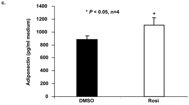
a. Quantitative real time RT-PCR measurement of adiponectin in cultured rat adult cardiomyocytes treated with rosiglitazone (Rosi, 10 μMol/L) at 3, 6 and 12 hour time-points, respectively. Data are expressed as mean±SEM, n=4, * p<0.05, **P<0.01. b. Western blot analyses of adiponectin normalized to actin in cultured adult rat cardiomyocytes treated with rosiglitazone (Rosi, 10 μMol/L, 24 hours). Data are expressed as mean±SEM, n=4, * p<0.01. Representative autographic images of Western blot results on adiponectin protein expression treated with or without rosiglitazone (Rosi). c. ELISA measurement of adiponectin concentration in media of cultured rat adult cardiomyocytes with or without rosiglitazone treatment (Rosi, 10 μMol/L, 24 hours). Data are expressed as mean±SEM, n=4, *P<0.01.
Figure 4. Effects of rosiglitazone treatment on adiponectin expression.

a. Quantitative real time PCR measurement of adipoR1 in cultured rat adult cardiomyocytes treated with rosiglitazone (Rosi, 10 μMol/L, 24 hours). Data are expressed as mean±SEM, n=6, *P<0.05. b. Quantitative real time PCR measurement of adipoR2 in cultured rat adult cardiomyocytes treated with rosiglitazone (Rosi, 10 μMol/L, 24 hours). Data are expressed as mean±SEM, n=6, *P<0.01.
Adiponectin enhances rates of FAO and glucose uptake in cultured ventricular cardiomyocytes via activation of AMPK activity
To investigate a role of adiponectin on energy metabolism in adult ventricular cardiomyocytes, we measured 14C Palmitate oxidation rates in cultured cardiomyocytes treated with or without globular adiponectin (2 μg/ml). Adiponectin pretreatment of cardiomyocytes produced substantially greater palmitate oxidation compared to control (Figure 5). We further examined whether adiponectin treatment alters glucose uptake in cultured cardiomyocytes. As anticipated, adiponectin treatment substantially increased basal glucose uptake and further increased insulin-stimulated glucose uptake in cultured cardiomyocytes (Figure 6). Therefore, adiponectin appears to be a positive regulator of both FAO and glucose uptake in cardiomyocytes presumably mediated by its two abundantly expressed receptors on cardiomyocytes. To investigate whether AMPK activation mediates adiponectin’s effects on energy metabolism in cardiomyocytes, we evaluated AMPK activity by measuring the phosphorylation state of one of its substrates ACC. As expected, cardiomyocytes treated with adiponectin demonstrated increased pACC without altering ACC protein levels compared with vehicle treated cardiomyocytes (Figure 7a). The increase in pACC levels peaked at 15 minutes after adiponectin treatment (Figure 7a). ACC activation was associated with a subsequent increase of MCD activity (Figure 7b) and a decrease in malonyl Co-A content (Figure 7b) in cultured cardiomyocytes treated with adiponectin. These results indicate that activation of AMPK may mediate the functional effects of adiponectin on augmentation of FAO and glucose uptake in cardiomyocytes.
Figure 5. Effects of adiponectin treatment on FAO in cultured rat cardiomyocytes.

Relative palmitate oxidation rates in adult rat cardiomyocytes with and without treatment of recombinant globular adiponectin (2 μg/ml) are shown. Palmitate oxidation rates in cardiomyocytes treated with adiponectin are expressed as percentage changes compared with control, which was set at 100%. Data are expressed as mean±SEM, n=4, *P<0.05.
Figure 6. Effects of adiponectin treatment on glucose uptake in cultured rat cardiomyocytes.

Glucose uptake in adult rat cardiomyocytes with and without treatment of globular adiponectin (2 μg/ml) was measured by the relative activities of 2-D-3H-glucose in cardiomyocytes. Specific activities of [3H]-2-deoxyglucose in cardiomyocytes with adiponectin and/or insulin treatments were expressed as percentage changes compared with those with vehicle control, which was set at 100%. Data are expressed as mean±SEM (n=6) and analyzed by two-way ANOVA. Adiponectin (A) or insulin (B) vs vehicle: *P<0.01. There is no interaction effect between adiponectin and insulin (C and D): P>0.05.
Figure 7.


a. Western blot analyses of pACC and ACC in rat cardiomyocytes treated with adiponectin (2 μg/ml) at different time points. The protein levels of pACC and ACC normalized to those of actin are shown. Data are expressed as mean±SEM, n=4, *P<0.01. b. MCD activity measured by spectrophotometric meathod in cardiomyocytes with and without treatment of globular adiponectin (2 μg/ml) for 30 minutes. Data are expressed as mean±SEM, n=4, *P<0.05. c). Malonyl CoA content measured by HPLC in adult rat cardiomyocytes (25×105 cells per flask) with and without treatment of globular adiponectin (2 μg/ml) for 30 minutes. Data are expressed as mean±SEM, n=5, *P<0.05.
Discussion
The present study demonstrates that adiponectin and its two receptors are expressed in adult ventricular cardiomyocytes and enhanced by PPARγ activation. Treatment of adiponectin activates AMPK activity and subsequently upregulates FAO and glucose uptake in adult cardiomyocytes.
Adiponectin has long been defined as an adipocyte-specific secretary protein with insulin-sensitizing effects on the liver and skeletal muscle [21–23]. It was reported that adiponectin is expressed in bone-forming cells [24] and its expression may be induced in cultured myotubes from skeletal muscle [25]. A recent report revealed that adiponectin can be detected from atrium-derived cardiomyocytes [4]. However, it remains unclear if adiponectin is expressed in ventricular cardiomyocytes, especially those from adult hearts. It also remains unclear on how the cardiac adiponectin expression is regulated and what are its roles in cardiomyocytes.
The relatively abundant expression of adiponectin in adult ventricular cardiomyocytes is quite a surprise. In this study, we utilized purified primary cardiomyocytes cultures being careful to exclude potential contamination by adipocytes to explore cardiac expression of adiponectin. Recently, a similar cardiac expression of leptin, another protein hormone that is involved in energy metabolic regulation, has been reported [26]. Adiponectin belongs to an adipokine superfamily. Another adipokine, TNFα, is also expressed and secreted from cardiomyocytes [27]. Interestingly, the globular domain of adiponectin displays highly similar homo-trimeric three-dimensional structures to that of TNFα, even though they have unrelated amino-acid sequences [28]. Therefore, adiponectin and TNFα may represent adipokines with important biological roles in cardiomyocytes. Recently, it was reported that adiponectin is localized at the periphery of damaged myocytes from myocardial and dilated cardiomyopathy patients [29]. Also, it has been shown that adiponectin treatment inhibits the progression of viral myocarditis through binding to the adiponectin receptor 1 in leptin-deficient mice [30]. Together, these findings and ours suggest that a local cardiac adiponectin system may represent a critical protective mechanism for the heart at pathological conditions.
Two adiponectin receptors, adipoR1 and adipoR2, that mediate the biological effects of adiponectin was identified recently [3]. AdipoR1 is a high-affinity receptor for globular adiponectin and a low-affinity receptor for the full-length ligand, whereas adipoR2 is an intermediate-affinity receptor for both forms of adiponectin [3]. AdipoR1 is abundantly expressed in skeletal muscle, whereas adipoR2 is predominantly expressed in the liver; therefore, only full-length adiponectin is active in the liver [3]. Since our findings indicate that both adipoR1 and adipoR2 are expressed in cardiomyocytes at levels comparable to those found in skeletal muscle and liver, respectively (Figure 2); thus, it is plausible to predict that cardiomyocytes should respond to adiponectin at various forms. This again emphasizes that adiponectin may play a pivotal role in regulating myocardial energy homeostasis. Accumulating evidence has shown that decreased levels of circulating adiponectin are associated with obesity, insulin resistance, Type 2 diabetes, and atherosclerosis, and that administration of adiponectin diminishes abnormalities associated with Metabolic Syndrome X [31]. Metabolic syndrome X is a major risk factor of cardiac dysfunction and heart failure [32]. Results from our current studies confirmed that adiponectin activates AMPK on cardiomyocytes, which in turn upregulates FAO and glucose uptake. Therefore, the cardiac adiponectin system appears to be essential for maintaining cardiac function in patients of Metabolic Syndrome X. The cardiac adiponectin described in the present study may exert its effects via autocrine or paracrine mechanisms. In this manner, even small amounts of adiponectin may exert a potent function on the cardiomyocytes. Alternatively, cardiomyocyte-derived adiponectin may serve as a reserved regulator of myocardial energy metabolism when circulating adiponectin becomes scarce in situations such as Metabolic Syndrome X. Further studies on the expression patterns of adiponectin and its receptors in myocardial tissue in obesity should be informative.
Our present results suggest that PPARγ might be one of the key determinants of myocardial energy homeostasis at least, in part, by regulating the expression of adiponectin and its receptors. Adiponectin promoter possesses a functional PPAR-responsive element (PPRE) and PPARγ/RXR heterodimer can directly bind to the PPRE and increase its promoter activity in cells [33]. PPARγ activation increases plasma levels of adiponectin in humans, while patients with dominant-negative PPARγ mutations exhibit substantially reduced adiponectin levels [34]. Contradictory results have been reported on whether PPARγ activation influences expression of adipoR1 and adipoR2 [35, 36]. Nonetheless, our studies support that adiponectin receptors are transcriptional targets of PPARγ in cardiac tissue. Thus, PPARγ activation could potentially affect adiponectin downstream signaling pathways. How PPARγ activation affects the expression of adiponectin receptors in cardiomyocytes remains unclear. However, we cannot exclude the possibility that they are regulated by the levels of adiponectin.
We demonstrated that the globular form of adiponectin augmented FAO and glucose uptake in cultured cardiomyocytes. Whether the full-length monomeric and/or multimeric forms of adiponectin also affect these or other biological properties of cardiomyocytes requires further evaluation. Given that adipoR2 is abundantly expressed in cardiomyocytes (Figure 2b), it is likely that full-length adiponectin may also influence cardiomyocyte function.
Malonyl-CoA is synthesized in the heart by ACC and is a potent endogenous inhibitor of mitochondrial FAO [37]. ACC is phosphorylated and inactivated by AMPK (see review [6]). In light of this, we hypothesized that adiponectin-mediated augmentation of FAO in cardiomyocytes is associated with AMPK activation and a concomitant increase in ACC phosphorylation and decrease in malonyl-CoA. Our data supported our hypothesis and were consistent with the findings of Yamauchi et al, which reveal a similar phenomenon in skeletal muscle and liver [2]. Conversely, another study reported earlier that globular adiponectin perfusion in isolated rabbit hearts increases myocardial FAO independent of AMPK activation [38]. This discrepancy may be due to different experimental approaches, animal species, in vitro cell culture versus ex vivo organ perfusion, as well as various time courses.
Taken together, the present study suggests that adiponectin may be an excellent candidate involved in the fine tuning of the heart’s responses to energetic needs under both physiological and pathological conditions. The cardiac adiponectin system may coordinate regulation of myocardial energy balance in response to systemic and local needs under physiologic and pathologic conditions. Therefore, adiponectin may be a potential therapeutic alternative in treating cardiac energetic anomalies.
Acknowledgments
This work was partially supported by grants from NIH (MBRS S06GM08248, 1R01HL085499 and 1R01HL084456) and a scientist development award from the American Heart Association national center. The investigation was conducted in a facility constructed with support from Research Facilities Improvement Grant #C06 RR- 1 C06 RR07571. We thank Dr. Bryan Slinker and Mr. Yong Liu for technical help on statistical analyses.
Footnotes
Publisher's Disclaimer: This is a PDF file of an unedited manuscript that has been accepted for publication. As a service to our customers we are providing this early version of the manuscript. The manuscript will undergo copyediting, typesetting, and review of the resulting proof before it is published in its final citable form. Please note that during the production process errors may be discovered which could affect the content, and all legal disclaimers that apply to the journal pertain.
References
- 1.Scherer PE, Williams S, Fogliano M, Baldini G, Lodish HF. A novel serum protein similar to C1q, produced exclusively in adipocytes. J Biol Chem. 1995 Nov 10;270(45):26746–9. doi: 10.1074/jbc.270.45.26746. [DOI] [PubMed] [Google Scholar]
- 2.Yamauchi T, Kamon J, Minokoshi Y, Ito Y, Waki H, Uchida S, et al. Adiponectin stimulates glucose utilization and fatty-acid oxidation by activating AMP-activated protein kinase. Nature medicine. 2002 Nov;8(11):1288–95. doi: 10.1038/nm788. [DOI] [PubMed] [Google Scholar]
- 3.Yamauchi T, Kamon J, Ito Y, Tsuchida A, Yokomizo T, Kita S, et al. Cloning of adiponectin receptors that mediate antidiabetic metabolic effects. Nature. 2003 Jun 12;423(6941):762–9. doi: 10.1038/nature01705. [DOI] [PubMed] [Google Scholar]
- 4.Pineiro R, Iglesias MJ, Gallego R, Raghay K, Eiras S, Rubio J, et al. Adiponectin is synthesized and secreted by human and murine cardiomyocytes. FEBS letters. 2005 Sep 26;579(23):5163–9. doi: 10.1016/j.febslet.2005.07.098. [DOI] [PubMed] [Google Scholar]
- 5.Shibata R, Ouchi N, Ito M, Kihara S, Shiojima I, Pimentel DR, et al. Adiponectin-mediated modulation of hypertrophic signals in the heart. Nature medicine. 2004 Dec;10(12):1384–9. doi: 10.1038/nm1137. [DOI] [PMC free article] [PubMed] [Google Scholar]
- 6.Hardie DG, Carling D. The AMP-activated protein kinase--fuel gauge of the mammalian cell? Eur J Biochem. 1997 Jun 1;246(2):259–73. doi: 10.1111/j.1432-1033.1997.00259.x. [DOI] [PubMed] [Google Scholar]
- 7.Kemp BE, Mitchelhill KI, Stapleton D, Michell BJ, Chen ZP, Witters LA. Dealing with energy demand: the AMP-activated protein kinase. Trends Biochem Sci. 1999 Jan;24(1):22–5. doi: 10.1016/s0968-0004(98)01340-1. [DOI] [PubMed] [Google Scholar]
- 8.Tian R, Musi N, D’Agostino J, Hirshman MF, Goodyear LJ. Increased adenosine monophosphate-activated protein kinase activity in rat hearts with pressure-overload hypertrophy. Circulation. 2001 Oct 2;104(14):1664–9. doi: 10.1161/hc4001.097183. [DOI] [PubMed] [Google Scholar]
- 9.Mitcheson JS, Hancox JC, Levi AJ. Cultured adult cardiac myocytes: future applications, culture methods, morphological and electrophysiological properties. Cardiovasc Res. 1998 Aug;39(2):280–300. doi: 10.1016/s0008-6363(98)00128-x. [DOI] [PubMed] [Google Scholar]
- 10.Shimoyama M, Hayashi D, Takimoto E, Zou Y, Oka T, Uozumi H, et al. Calcineurin plays a critical role in pressure overload-induced cardiac hypertrophy. Circulation. 1999 Dec 14;100(24):2449–54. doi: 10.1161/01.cir.100.24.2449. [DOI] [PubMed] [Google Scholar]
- 11.Kim YC, Gomez FE, Fox BG, Ntambi JM. Differential regulation of the stearoyl-CoA desaturase genes by thiazolidinediones in 3T3-L1 adipocytes. J Lipid Res. 2000 Aug;41(8):1310–6. [PubMed] [Google Scholar]
- 12.Nagao K, Inoue N, Wang YM, Yanagita T. Conjugated linoleic acid enhances plasma adiponectin level and alleviates hyperinsulinemia and hypertension in Zucker diabetic fatty (fa/fa) rats. Biochem Biophys Res Commun. 2003 Oct 17;310(2):562–6. doi: 10.1016/j.bbrc.2003.09.044. [DOI] [PubMed] [Google Scholar]
- 13.Mitsumoto Y, Klip A. Development regulation of the subcellular distribution and glycosylation of GLUT1 and GLUT4 glucose transporters during myogenesis of L6 muscle cells. J Biol Chem. 1992 Mar 5;267(7):4957–62. [PubMed] [Google Scholar]
- 14.Inukai K, Nakashima Y, Watanabe M, Takata N, Sawa T, Kurihara S, et al. Regulation of adiponectin receptor gene expression in diabetic mice. Am J Physiol Endocrinol Metab. 2005 May;288(5):E876–82. doi: 10.1152/ajpendo.00118.2004. [DOI] [PubMed] [Google Scholar]
- 15.Caminos JE, Nogueiras R, Gallego R, Bravo S, Tovar S, Garcia-Caballero T, et al. Expression and regulation of adiponectin and receptor in human and rat placenta. J Clin Endocrinol Metab. 2005 Jul;90(7):4276–86. doi: 10.1210/jc.2004-0930. [DOI] [PubMed] [Google Scholar]
- 16.Cheng L, Ding G, Qin Q, Xiao Y, Woods D, Chen YE, et al. Peroxisome proliferator-activated receptor [delta] activates fatty acid oxidation in cultured neonatal and adult cardiomyocytes. Biochemical and Biophysical Research Communications. 2004 Jan 9;313(2):277–86. doi: 10.1016/j.bbrc.2003.11.127. [DOI] [PubMed] [Google Scholar]
- 17.Cheng L, Ding G, Qin Q, Huang Y, Lewis W, He N, et al. Cardiomyocyte-restricted peroxisome proliferator-activated receptor-delta deletion perturbs myocardial fatty acid oxidation and leads to cardiomyopathy. Nature medicine. 2004 Nov;10(11):1245–50. doi: 10.1038/nm1116. [DOI] [PubMed] [Google Scholar]
- 18.King MT, Reiss PD. Separation and measurement of short-chain coenzyme-A compounds in rat liver by reversed-phase high-performance liquid chromatography. Anal Biochem. 1985 Apr;146(1):173–9. doi: 10.1016/0003-2697(85)90412-9. [DOI] [PubMed] [Google Scholar]
- 19.Saddik M, Gamble J, Witters LA, Lopaschuk GD. Acetyl-CoA carboxylase regulation of fatty acid oxidation in the heart. J Biol Chem. 1993 Dec 5;268(34):25836–45. [PubMed] [Google Scholar]
- 20.Kim YS, Kolattukudy PE. Malonyl-CoA decarboxylase from the uropygial gland of waterfowl: purification, properties, immunological comparison, and role in regulating the synthesis of multimethyl-branched fatty acids. Arch Biochem Biophys. 1978 Oct;190(2):585–97. doi: 10.1016/0003-9861(78)90314-4. [DOI] [PubMed] [Google Scholar]
- 21.Yamamoto K, Ohki R, Lee RT, Ikeda U, Shimada K. Peroxisome proliferator-activated receptor gamma activators inhibit cardiac hypertrophy in cardiac myocytes. Circulation. 2001;104(14):1670–5. doi: 10.1161/hc4001.097186. [DOI] [PubMed] [Google Scholar]
- 22.Yamauchi T, Kamon J, Waki H, Terauchi Y, Kubota N, Hara K, et al. The fat-derived hormone adiponectin reverses insulin resistance associated with both lipoatrophy and obesity. Nature medicine. 2001 Aug;7(8):941–6. doi: 10.1038/90984. [DOI] [PubMed] [Google Scholar]
- 23.Berg AH, Combs TP, Du X, Brownlee M, Scherer PE. The adipocyte-secreted protein Acrp30 enhances hepatic insulin action. Nature medicine. 2001 Aug;7(8):947–53. doi: 10.1038/90992. [DOI] [PubMed] [Google Scholar]
- 24.Berner HS, Lyngstadaas SP, Spahr A, Monjo M, Thommesen L, Drevon CA, et al. Adiponectin and its receptors are expressed in bone-forming cells. Bone. 2004 Oct;35(4):842–9. doi: 10.1016/j.bone.2004.06.008. [DOI] [PubMed] [Google Scholar]
- 25.Delaigle AM, Jonas JC, Bauche IB, Cornu O, Brichard SM. Induction of adiponectin in skeletal muscle by inflammatory cytokines: in vivo and in vitro studies. Endocrinology. 2004 Aug 19; doi: 10.1210/en.2004-0503. [DOI] [PubMed] [Google Scholar]
- 26.Purdham DM, Zou MX, Rajapurohitam V, Karmazyn M. Rat heart is a site of leptin production and action. Am J Physiol Heart Circ Physiol. 2004 Dec;287(6):H2877–84. doi: 10.1152/ajpheart.00499.2004. [DOI] [PubMed] [Google Scholar]
- 27.Meldrum DR. Tumor necrosis factor in the heart. Am J Physiol. 1998 Mar;274(3 Pt 2):R577–95. doi: 10.1152/ajpregu.1998.274.3.R577. [DOI] [PubMed] [Google Scholar]
- 28.Shapiro L, Scherer PE. The crystal structure of a complement-1q family protein suggests an evolutionary link to tumor necrosis factor. Curr Biol. 1998 Mar 12;8(6):335–8. doi: 10.1016/s0960-9822(98)70133-2. [DOI] [PubMed] [Google Scholar]
- 29.Takahashi T, Saegusa S, Sumino H, Nakahashi T, Iwai K, Morimoto S, et al. Adiponectin, T-cadherin and tumour necrosis factor-alpha in damaged cardiomyocytes from autopsy specimens. J Int Med Res. 2005 Mar-Apr;33(2):236–44. doi: 10.1177/147323000503300212. [DOI] [PubMed] [Google Scholar]
- 30.Takahashi T, Saegusa S, Sumino H, Nakahashi T, Iwai K, Morimoto S, et al. Adiponectin replacement therapy attenuates myocardial damage in leptin-deficient mice with viral myocarditis. J Int Med Res. 2005 Mar-Apr;33(2):207–14. doi: 10.1177/147323000503300208. [DOI] [PubMed] [Google Scholar]
- 31.Matsuzawa Y, Funahashi T, Kihara S, Shimomura I. Adiponectin and metabolic syndrome. Arterioscler Thromb Vasc Biol. 2004 Jan;24(1):29–33. doi: 10.1161/01.ATV.0000099786.99623.EF. [DOI] [PubMed] [Google Scholar]
- 32.Kenchaiah S, Evans JC, Levy D, Wilson PW, Benjamin EJ, Larson MG, et al. Obesity and the risk of heart failure. N Engl J Med. 2002 Aug 1;347(5):305–13. doi: 10.1056/NEJMoa020245. [DOI] [PubMed] [Google Scholar]
- 33.Iwaki M, Matsuda M, Maeda N, Funahashi T, Matsuzawa Y, Makishima M, et al. Induction of adiponectin, a fat-derived antidiabetic and antiatherogenic factor, by nuclear receptors. Diabetes. 2003 Jul;52(7):1655–63. doi: 10.2337/diabetes.52.7.1655. [DOI] [PubMed] [Google Scholar]
- 34.Combs TP, Wagner JA, Berger J, Doebber T, Wang WJ, Zhang BB, et al. Induction of adipocyte complement-related protein of 30 kilodaltons by PPARgamma agonists: a potential mechanism of insulin sensitization. Endocrinology. 2002 Mar;143(3):998–1007. doi: 10.1210/endo.143.3.8662. [DOI] [PubMed] [Google Scholar]
- 35.Chinetti G, Zawadski C, Fruchart JC, Staels B. Expression of adiponectin receptors in human macrophages and regulation by agonists of the nuclear receptors PPARalpha, PPARgamma, and LXR. Biochem Biophys Res Commun. 2004 Jan 30;314(1):151–8. doi: 10.1016/j.bbrc.2003.12.058. [DOI] [PubMed] [Google Scholar]
- 36.Kaltenbach S, Staiger H, Weisser M, Haas C, Stumvoll M, Machicao F, et al. Adiponectin receptor gene expression in human skeletal muscle cells is not regulated by fibrates and thiazolidinediones. Int J Obes (Lond) 2005 Jul;29(7):760–5. doi: 10.1038/sj.ijo.0802957. [DOI] [PubMed] [Google Scholar]
- 37.Lopaschuk GD, Witters LA, Itoi T, Barr R, Barr A. Acetyl-CoA carboxylase involvement in the rapid maturation of fatty acid oxidation in the newborn rabbit heart. J Biol Chem. 1994 Oct 14;269(41):25871–8. [PubMed] [Google Scholar]
- 38.Onay-Besikci A, Altarejos JY, Lopaschuk GD. gAd-globular head domain of adiponectin increases fatty acid oxidation in newborn rabbit hearts. J Biol Chem. 2004 Oct 22;279(43):44320–6. doi: 10.1074/jbc.M400347200. [DOI] [PubMed] [Google Scholar]